An archive of images by HubLE international community of early- and mid-career basic and clinical scientists in the field of musculoskeletal research.
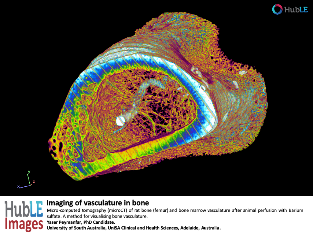
Imaging of vascular in bone.
Click image to enlarge
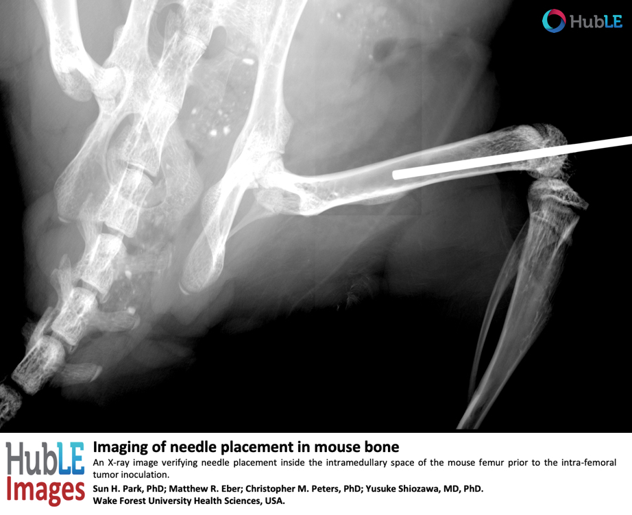
Imaging of needle placement in mouse bone
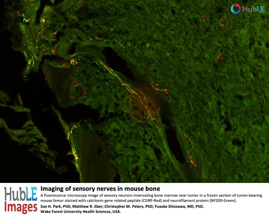
Imaging of sensory nerves in mouse bone
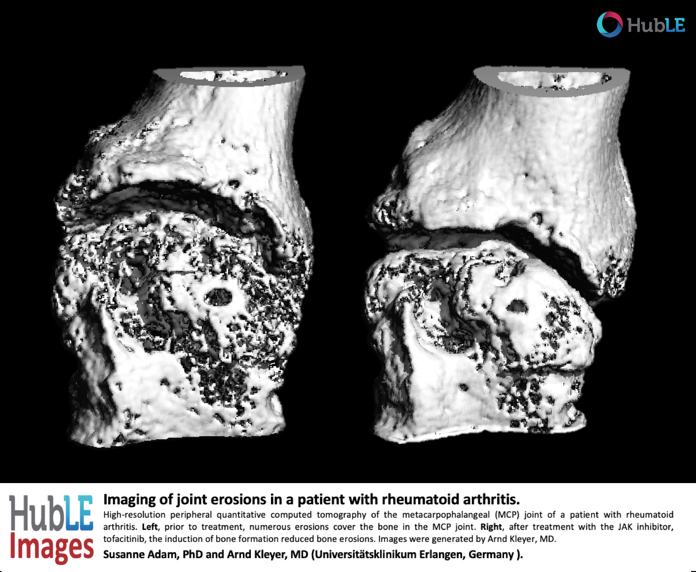
Joint erosions in a patient with rheumatoid arthritis.
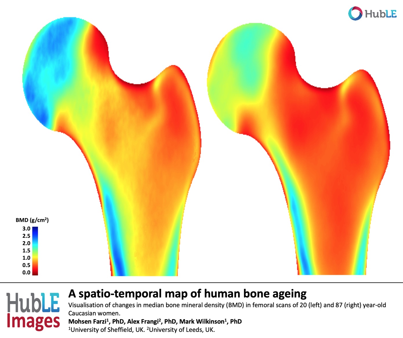
A spatio-temporal map of human bone ageing
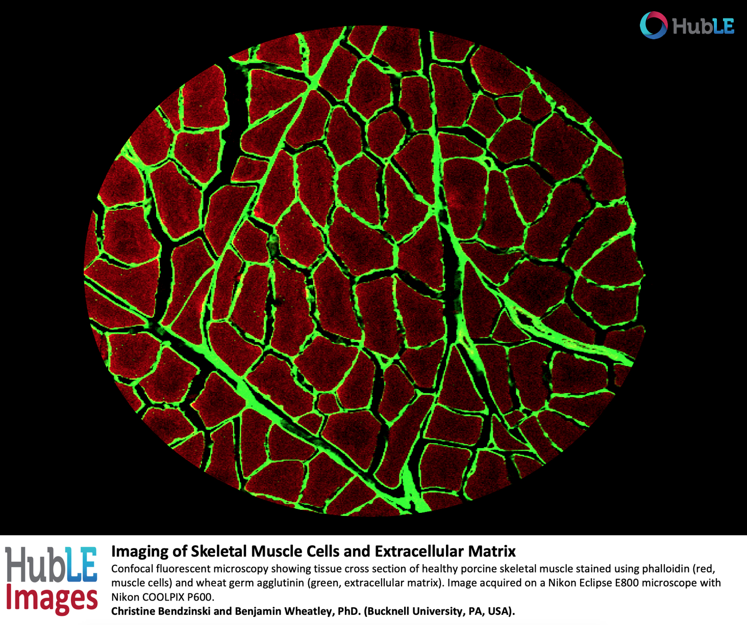
Imaging of Skeletal Muscle Cells and Extracellular Matrix
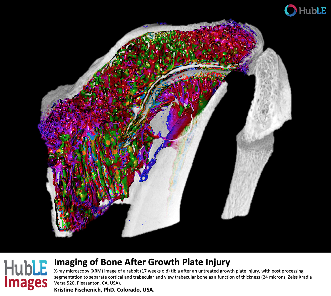
Imaging of Bone After Growth Plate Injury
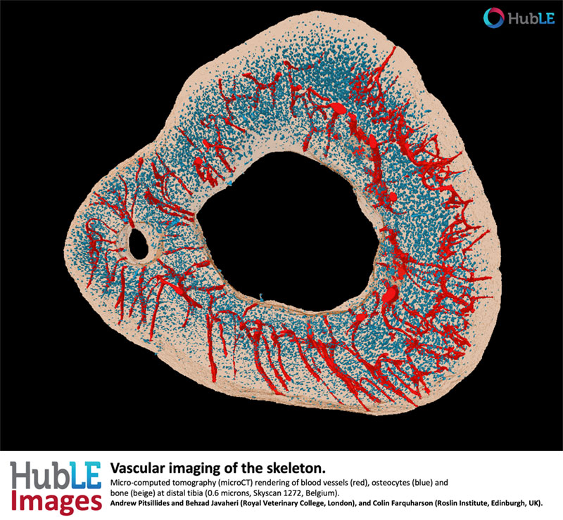
Vascular imaging of the skeleton
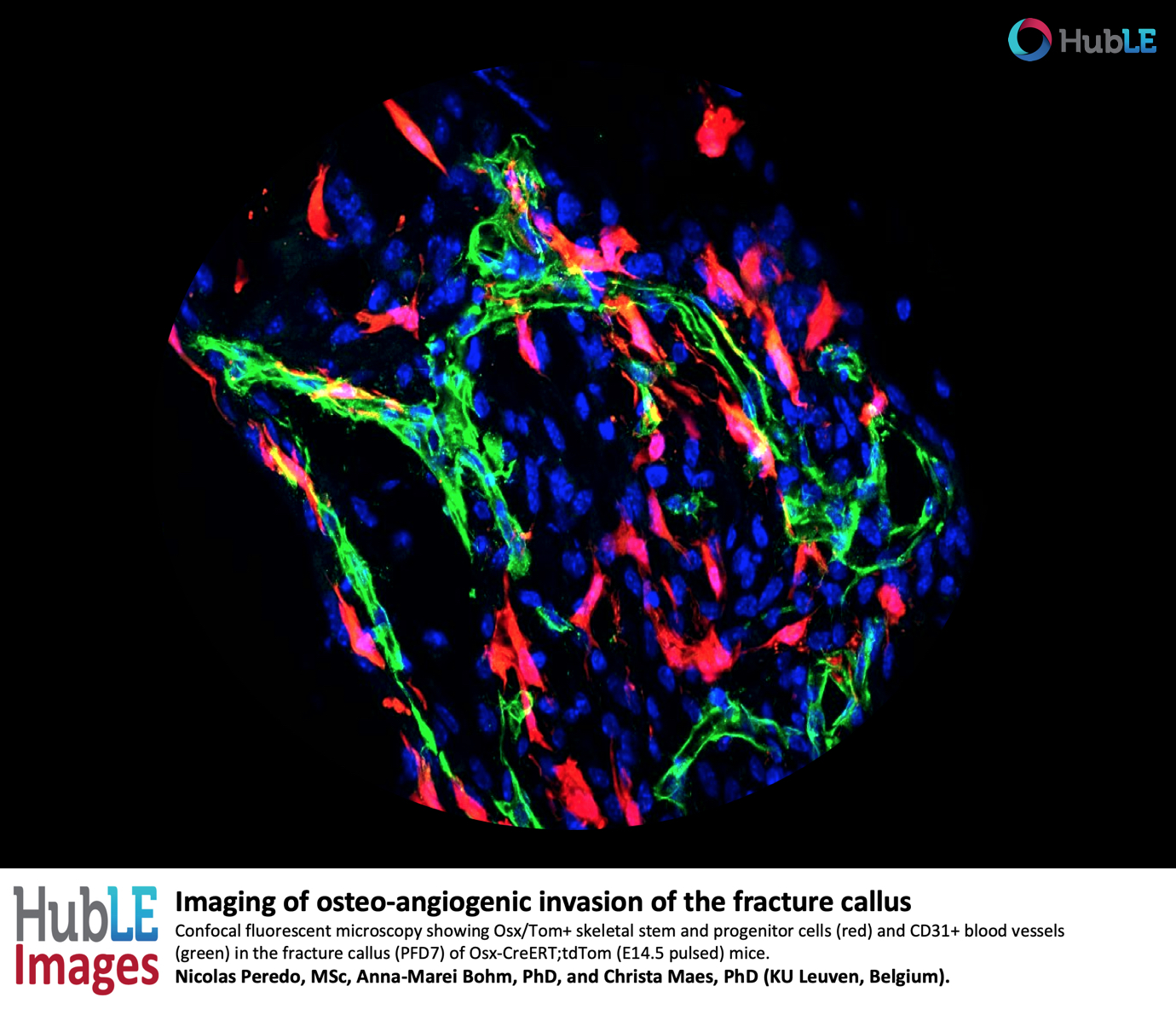
Imaging of osteo-angiogenic invasion of the fracture callus
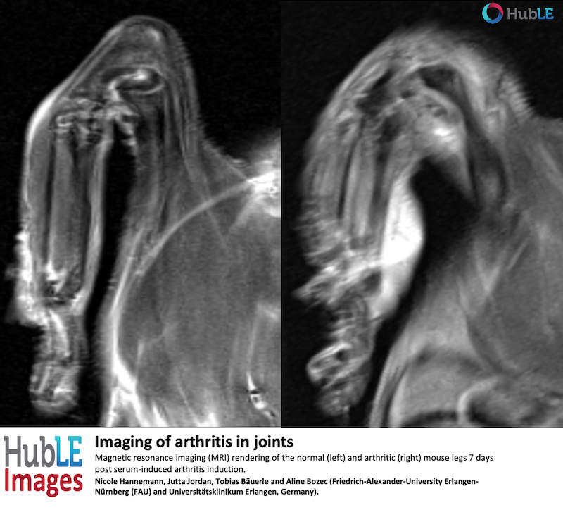
Imaging of arthritis in joints
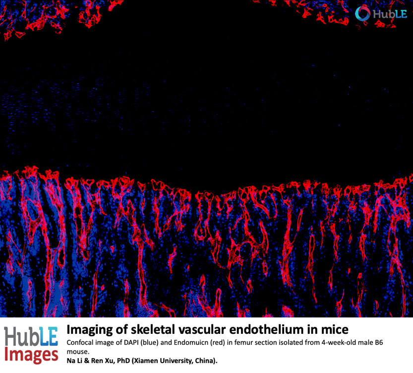
Skeletal vascular endothelium
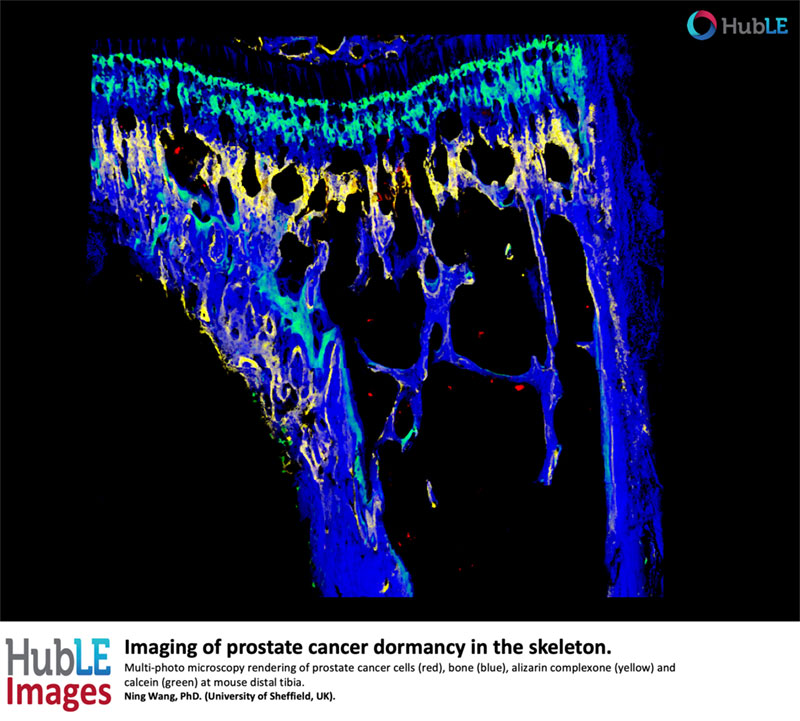
Imaging of prostate cancer dormancy
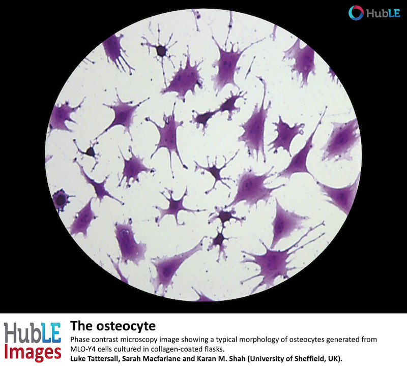
The osteocyte
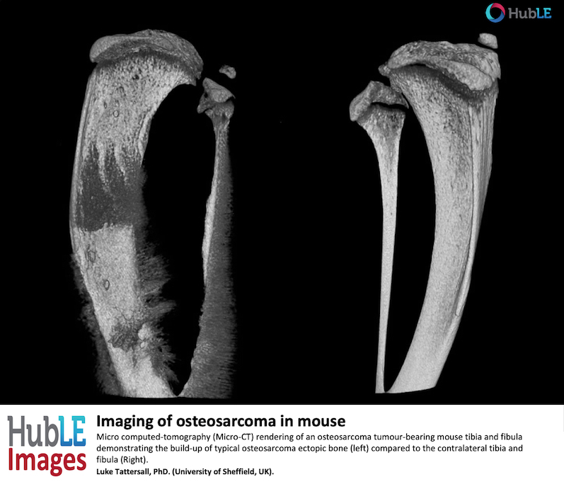
Imaging of osteosarcoma in mouse
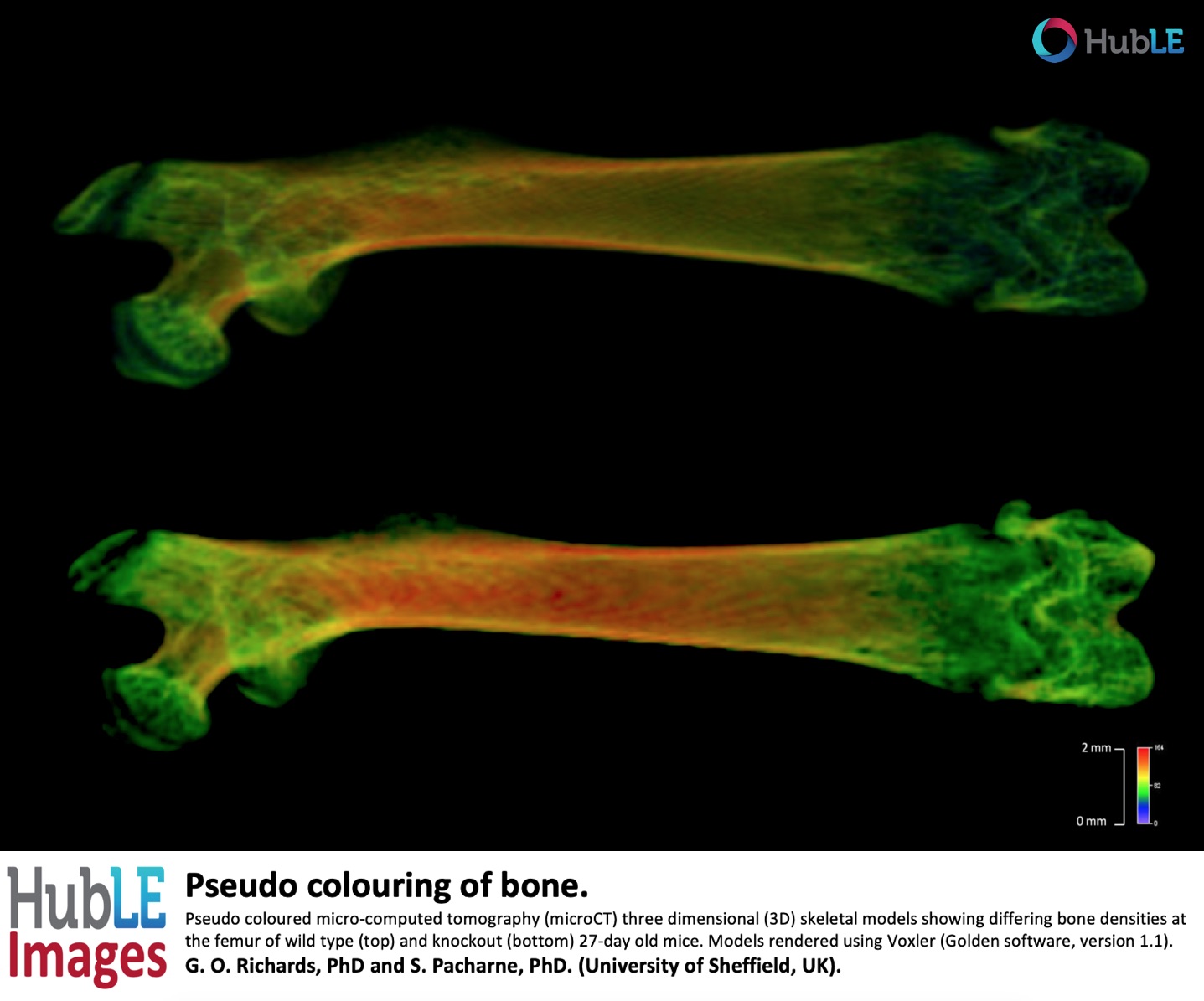
Pseudo colouring of bone.
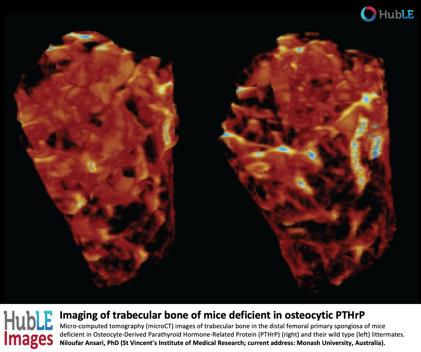
Imaging of trabecular bone of mice deficient in osteocytic PTHrP
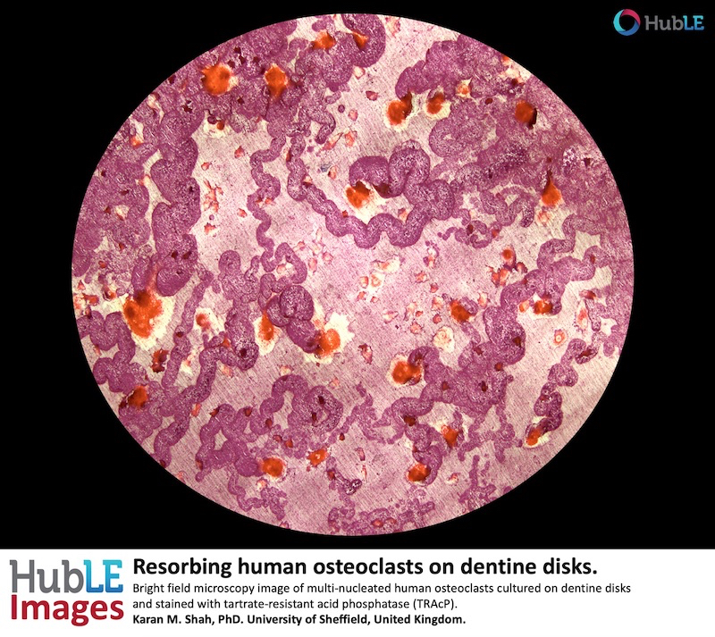
Resorbing human osteoclast
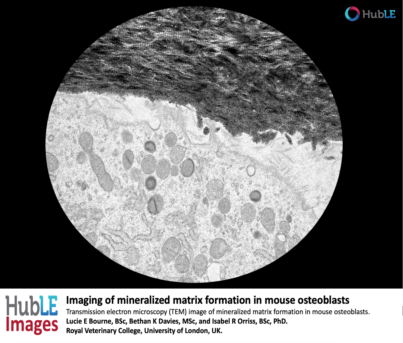
Imaging of mineralised matrix formation in mouse osteoblasts
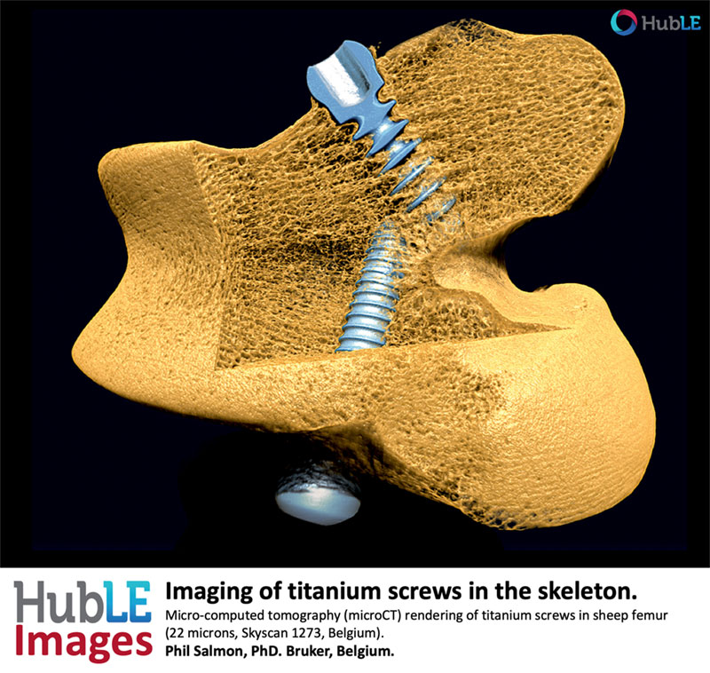
Titanium screws in the skeleton
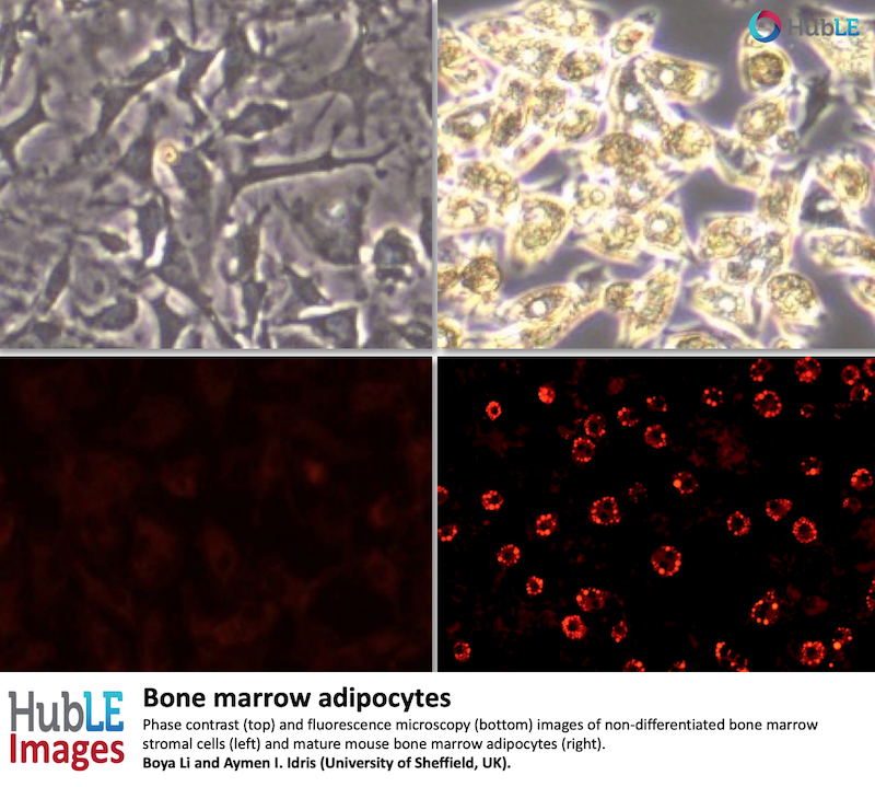
Bone marrow adipocytes
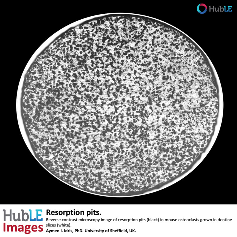
Resorption pits
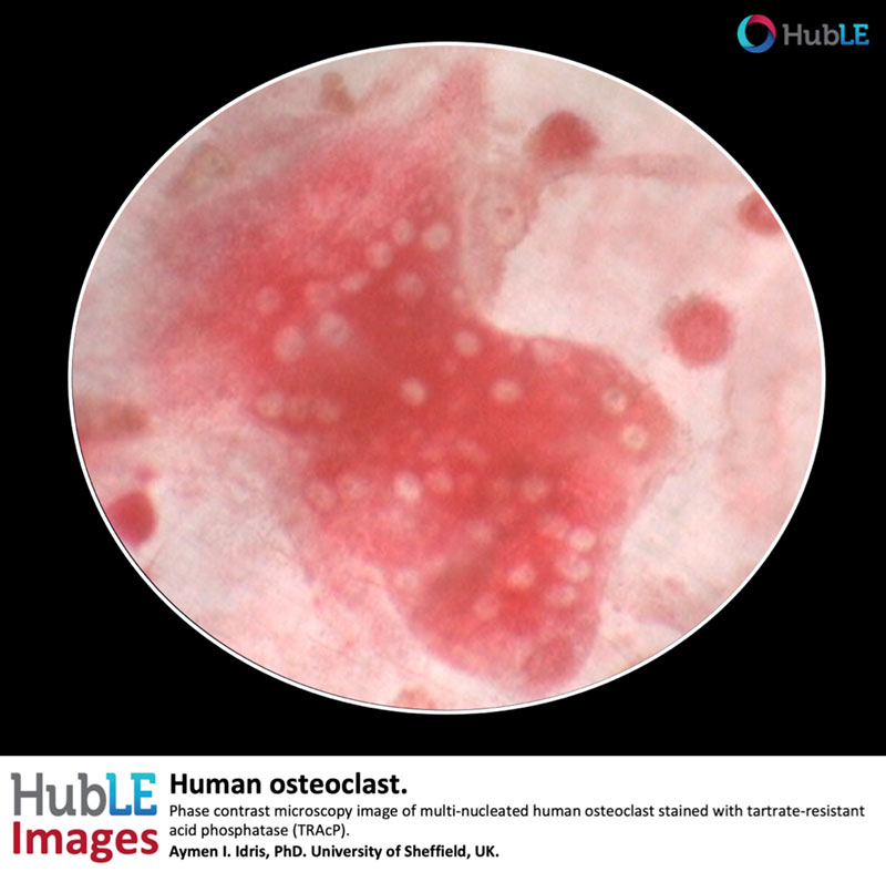
Human osteoclast
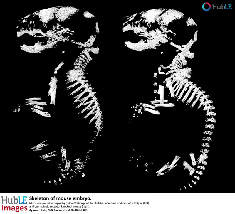
Skeleton of mouse embryo